

Having reached the level of excellence in line with the advancements in technology since 1970’s, when they were first used, bronchoscopes saw the most significant breakthrough in early 2000’s. The ultrasonography device, which had already been in use for medical purposes for about 70 years at that time, was combined with the bronchoscope, introducing endobronchial ultrasonography (EBUS). The reason why this was such a notable development was that EBUS could now be used not only in the bronchial tree but also in mediastinum, the medium section of thorax, and extreme ends of lungs.
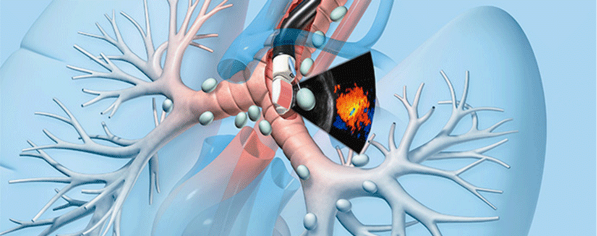
EBUS essentially comprises a flexible bronchoscope and an ultrasonography device. The ultrasonography probe may be affixed to the tip of the bronchoscope or it may be advanced through the channel inside the bronchoscope. Although only brief descriptions are made here, this revolutionary innovation actually broke new grounds in the field of pulmonary diseases. Thanks to EBUS, some inpatient diagnostic surgical interventions previously performed under general anesthesia, which many patients had to undergo, are no longer necessary.
Used mostly for the purpose of diagnosing and staging pulmonary cancer, EBUS is also utilized in diagnosis of sarcoidosis, lymphoma and various other types of cancer and diseases. It can be performed under general or local anesthesia + sedation without hospitalization.
A correct staging study is of utmost importance in configuring an accurate mode of therapy for lung cancer. Finding out whether tumoral spread is present in mediastinal lymph nodes is the determining factor for treatment of lung cancer patients who do not present with any organ metastasis. With EBUS, ultrasonographic view of these lymph nodes can be obtained from within bronchi and specimens can be collected for microscopic examination. This process takes approximately 15-30 minutes and collected specimens can be evaluated by a pathologist in the same room while the procedure is still in progress, which effectively reduces the overall time required for a diagnosis while enhancing diagnostic value of the entire investigation. Up to 95% of cases benefit from accurate staging, thanks to EBUS. A high rate of non-cancer patients can also be diagnosed. In rare cases where diagnosis may not be possible with EBUS, surgical methods like mediastinoscopy or thoracoscopy can be preferred.
Another noteworthy feature of endobronchial ultrasonography is its low risk of complication. The ability to conduct real-time imaging while collecting a biopsy sample is another major advantage. This enables visualization of vascular structures and prevents potential injuries.