

Mediastinum and Mediastinal Masses
The area behind our two lungs containing the heart, main arteries, thymus gland and esophagus is called the “mediastinum”. It is divided into three main anatomic regions: Anterior, middle and posterior mediastinum. Varying among patients of different age groups and genders, anterior mediastinum may house tumors of the thymus gland, lymphomas on lymph glands and germ-cell tumors. Tumors of nerve tissues are more commonly seen in posterior mediastinum. Any such case can be accurately diagnosed by taking into consideration the patient’s age, gender, symptoms, blood readings, computerized tomography images and uptake patterns in PET/CT scan. As with every other type of tumor, effective treatment depends on accurate diagnosis and staging. Some mediastinal tumors require surgical intervention or chemotherapy, while others should be treated with both approaches.
Thymoma
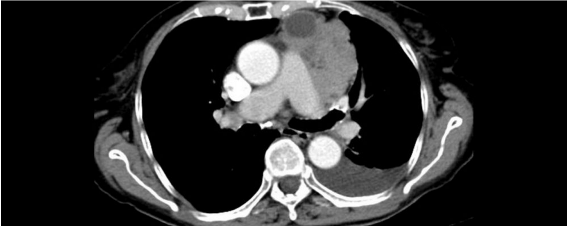
Thymoma is one of the most common types of tumor encountered in anterior mediastinum. It originates from the thymus gland. Even though it may not trigger any symptoms, it might also be present along with numerous other diseases. Most of the time it accompanies myasthenia gravis, which is a muscular disease. Surgery is the first option in cases of thymoma without metastasis over peripheral organs. If metastasis is present, however, chemotherapy should precede surgery. Surgical intervention options vary in accordance with the degree of tumoral spread. Thoracoscopic and robotic surgeries are successfully performed at our clinic for excision of smaller tumors at earlier stages of disease. These methods provide an effective solution without having to saw off the sternum, which should only be considered for larger-scale metastatic thymoma cases due to the associated pain and cosmetic concerns. Additional radiotherapy or chemotherapy, albeit seldom, may be necessary after surgery. Upon completion of the entire treatment, patients should be followed with chest tomography findings in specific intervals.