

Being the most commonly encountered type of cancer among the male population, lung cancer keeps growing more and more in prominence also among the female population due to increasing scope of smoking habit. Accounting for the highest number of cancer-related deaths on a global scale, it claimed 1.61 million lives in 2008. The big majority of lung cancer cases originate from emerging countries. This most possibly stems from widespread smoking and adverse working conditions present in such regions.
Despite its reputation as one of the most common types of cancer, it is also the only preventable type, considering the fact that it largely develops secondary to smoking. Various studies have proven that while its prominence is directly related to smoking, countries that have launched anti-smoking campaigns have managed to decrease not only the number of smokers but also incidence rate of lung cancer.
Risk Factors
A wide range of risk factors are associated with lung cancer, smoking being the most significant one, as it induces 80-85% of all lung cancer cases. The cause-effect relation between smoking and squamous cell lung cancer as well as small-cell lung cancer is particularly well-defined. Daily smoking habits and term of actual addiction are directly proportional to risk of incidence, which is to say frequent smokers face increased risk in comparison to non-smokers or to those who smoke less.
In addition to smoking, risk factors such as exposure to some metals and chemical substances, coming in contact with asbestos or radon gas, genetic factors, known history of specific diseases and dietary habits also play a role in lung cancer. Individuals who have a familial history of lung cancer, for instance, are slightly more susceptible to it than others, more if it is coupled with smoking. Tuberculosis in the lungs, interstitial fibrosis, bullous emphysema and obliteration of tissues or scar tissues resulting from some other pulmonary diseases might lay the groundwork for onset of cancer. Exposure to asbestos, the most common risk factor for mesothelioma (pleural cancer), also aggravates the risk of incidence.
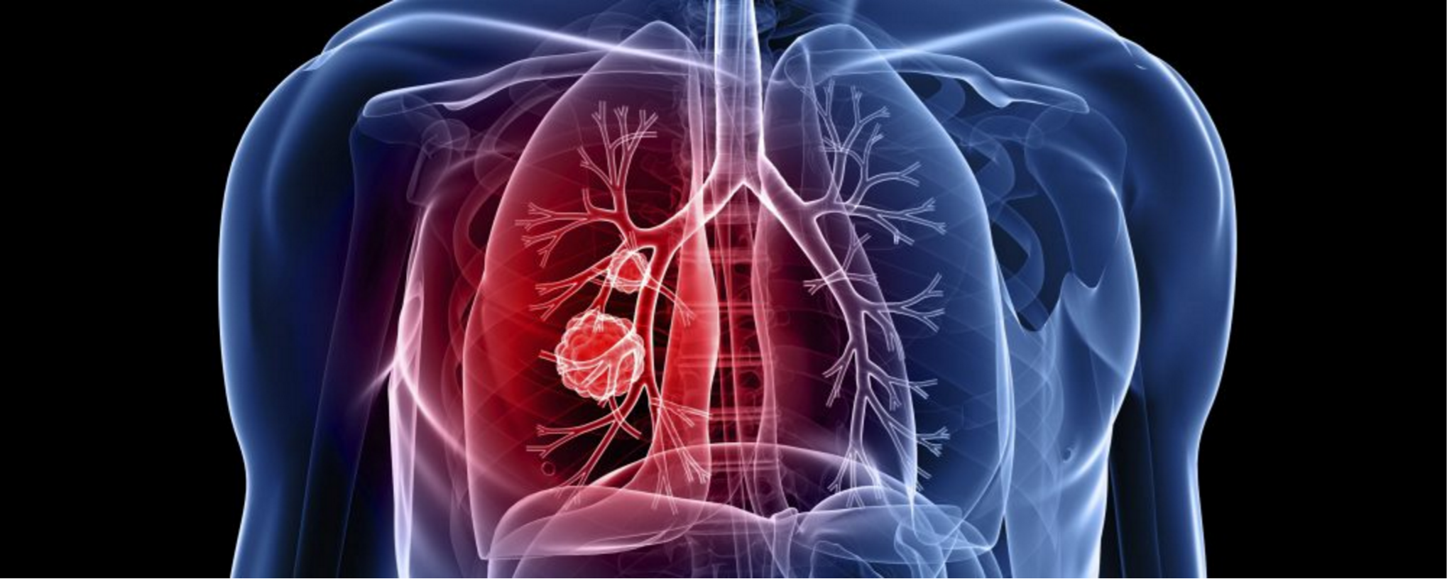
Classification of Lung Cancer by Cell Types
Lung cancer is classified in 2 basic categories in accordance with type of affected cells: Small cell and non-small cell. Non-small cell lung cancer includes the subgroups of squamous cell, large cell and adenocarcinoma. Course and treatment of small cell and non-small cell lung cancer differ in some aspects. Incidence rate of small cell lung cancer is 15-20% percent among all cases. Squamous cell cancer, a subgroup of non-small cell lung cancer, is one of the most common subtypes in our country and mainly occurs due to smoking. Although it is in the same category, incidence of adenocarcinoma is less frequent in our country, whereas it is the most prominent type of lung cancer in the USA. However, it is also a fact that incidence rate of adenocarcinoma has been increasing in our country over the years. Squamous cell cancer and small cell cancer occupy mostly central regions of the lung, i.e. main bronchi and lobar bronchi. Adenocarcinoma, on the other hand, mostly originates from peripheral areas of the lung.
Symptoms
Symptoms of lung cancer can be categorized as local and non-pulmonary symptoms:
Local symptoms stem directly from the tumor localized in the lung and its metastases over local lymph glands. These include coughing; expectoration; dyspnea; pain in the chest, shoulders, arms or back; hemoptysis; hoarseness; swelling in the face and neck; and wheezing. Regardless, most symptoms will not be visible during early stages of disease. Even though coughing is an early symptom in most cases, the big majority of patients tend not to consult a physician, as they associate coughing with their smoking habit. Therefore, seeing a specialized physician is definitely recommended in case of long-lasting coughs (ongoing for more than 3 weeks) or changes in existing coughing problem, such as discharge of blood and phlegm after a cough.
Non-pulmonary symptoms of lung cancer may be triggered either by tumoral metastases over other organs or some immunological and hormonal substances secreted by the tumor. Metastatic symptoms are organ-specific: Bone metastases manifest themselves with pain, while brain metastasis is accompanied with compromised consciousness, convulsive seizures and disrupted visual acuity. Weight loss, malaise, loss of strength and febrility may also be observed in addition to metastatic symptoms in most cases. Clubbing of the fingers, dermal lesions, neurological disorders and negative blood readings may be caused by tumoral secretions too, especially in cases of small cell lung cancer.
Diagnosis and Staging
After hearing the anamnesis of a patient who presents with the aforementioned symptoms and findings and conducting physical examination, the first step to be taken is obtaining a standard chest x-ray. Using this imaging technique, conditions like tumoral infections, pleuresis (fluid accumulation) and atelectasis (collapse of the lung) can be detected in most cases. If other tumor-associated symptoms are observed in chest x-ray, the next step is usually conducting a computerized tomography examination, which not only provides detailed information about the lesion at hand, but also visualizes lesions that are otherwise too small for chest x-ray to reveal. Following standard chest x-ray and computerized tomography examinations, local spread of the disease and localization can be identified. A physician will thus be able to decide on a method of biopsy for the purpose of attaining a final diagnosis. For example, performing bronchoscopy is essential for both diagnosis and staging of cases with surgical considerations and tumors with central localization. A biopsy sample can be harvested from tumors in more distal areas of the lung by traversing the cutaneous and subcutaneous layers with a needle under guidance of ultrasonography or computerized tomography. Moreover, recent technological breakthroughs have introduced endobronchial ultrasonography and navigation-assisted bronchoscopy methods, which make it possible to reach farther areas of the lung with bronchoscopy. After visualizing the tumor under chest x-ray and computerized tomography, staging studies should be initiated once a diagnosis specifying cell type has been obtained through methods like cytological analysis of phlegm and pleural fluid, bronchoscopy or needle biopsy. Staging of lung cancer is of critical importance in terms of determining the course of disease and suitable route of treatment, and should thus be definitely carried out.
Small cell lung cancer is evaluated in 2 stages: Limited and extensive. Disease is limited to one side of the chest without spread over the other lung or other organs in limited stage. In extensive stage, cancer has metastasized over non-pulmonary organs or the other lung. As treatment differs in limited and extensive stages, performing at least cranial tomography and MRI, bone scintigraphy and upper abdominal tomography or ultrasonography is essential to look into remote organ metastases in cases diagnosed with small cell lung cancer.
Taking into consideration that surgical intervention is the most effective mode of treatment for earlier stages of non-small cell lung cancer, staging of this subtype requires more detailed investigation than small cell lung cancer. Staging studies conducted for this purpose are in line with the TNM system. T is used to describe various features such as tumor size, relation to other tissues and organs and bronchoscopic appearance. N signifies the presence or lack of local or remote metastasis over lymph nodes. M is related to remote organ metastasis, while M0 cases do not exhibit metastasis.
Staging of lung cancer is ensured by employing primarily imaging and bronchoscopic methods and then surgical means, if deemed necessary. Imaging techniques like computerized tomography, cranial MRI and the more recent PET/CT as well as minimally invasive methods like bronchoscopy and endobronchial ultrasonography are used for staging. It is possible to reveal spread of lung cancer over lymph nodes within the thorax or organs and areas other than the lungs, if any, using PET and PET/CT studies. It should, however, be noted that lymph nodes within thorax which may appear suspicious for cancer in PET/CT should definitely be verified with one of the various biopsy methods. This is essentially important, because commonly encountered diseases in our country such as tuberculosis, other infectious diseases and non-cancer diseases like sarcoidosis can also be detected using PET/CT. Endobronchial ultrasonography is the foremost method recommended to collect biopsy samples from enlarged intrathoracic lymph nodes identified in PET/CT. It is a highly reliable and painless bronchoscopic method that can be practiced on outpatient basis without hospitalization. It offers more than 90% precision in staging. Surgical staging methods such as mediastinoscopy and thoracoscopy are also utilized in rare cases where endobronchial bronchoscopy does not yield a result.
Treatment
Following diagnosis and staging, a suitable mode of therapy should be considered. Lung cancer, in that sense, requires a multidisciplinary approach. This is to say treatment and follow-up of lung cancer should be outlined and performed jointly by physicians of pulmonary medicine, thoracic surgery, radiology, pathology and oncology.
Patients for whom surgical intervention is considered should preoperatively be evaluated by a physician of pulmonary medicine in order to prepare the patient for such intervention, determine possible risks and take risk mitigation measures. Preoperative evaluation involves physical examination and respiratory function tests. After this initial evaluation and reaching the conclusion that the patient’s pulmonary function is adequate, the patient is referred for surgery. On the other hand, findings indicating restricted respiratory function, COPD or other dormant pulmonary/cardiac diseases mean the case should be evaluated with cardiopulmonary exercise tests as well. Regardless of pulmonary function test results, ensuring that all patients quit smoking is essential. This effectively minimizes postoperative risks and slows down the rate at which disease progresses.
Treatment of lung cancer varies in accordance with its stages. A treatment program is available for each stage, and customized options are also presented to individual patients. If cancer is limited to only one lung, for instance, surgical intervention is prioritized to attain local control. Depending on characteristics of the tumor, chemotherapy and/or radiotherapy may be initiated postoperatively in some cases. In case the disease has advanced locally, exhibiting limited presence only in one lung but also having spread over lymph nodes outside its own margins, it is also possible to detect this prior to an operation, which then requires systemic chemotherapy followed by surgery, provided that local control and convenient circumstances have been attained. If the disease has metastasized out of one lung, it should be considered a systemic disease rather than one that needs to be controlled locally. In that case, therapy comprises chemotherapy and radiotherapy delivered either simultaneously or consecutively. In the end, different approaches may be adopted depending on patient-specific stage of disease, tumoral localization and pathological details. It is worth noting on this occasion that we make our decisions relevant to each non-standard case and patient who has undergone surgery at our weekly multidisciplinary oncology council meeting, which includes physicians from the clinics of pulmonary medicine, thoracic surgery, radiology, nuclear medicine, pathology, medical oncology and radiation oncology.